What is the name of the sponge? Sponges type classes. Internal structure of representatives
Despite the variety of body shapes and intricacy of appearance, all known sponges are divided into three main types based on their external shape, which received the following names: ascon, sicon and leucon.
Askon- the most primitive form of sponge body structure.
Asconoids have a small goblet-shaped or sac-shaped body, the base of which is fixed to the substrate. In the upper part of the body there is an opening - an opening, sometimes called the osculum.
Sponges - description, types, characteristics, nutrition, examples and classification
The body of such sponges is formed by two layers of cells - outer and inner, between which there is a gelatinous substance - mesoglea, consisting of cells of different structure and purpose. The outer layer of cells ("skin") is composed of flat cells - pinacocytes, which form the covering epithelium that separates the mesoglea from the external environment.
Among the pinacocytes there are larger cells - porocytes, which have an internal canal that opens outward and provides a connection between the inside of the sponge and the external environment.
The inner layer of the body is lined with collar cells - choanocytes, which have an elongated shape and a flagellum, the base of which is fixed in a plasmatic collar, which has the appearance of a funnel, open towards the internal cavity of the sponge.
Mesoglea contains immobile stellate cells (collencytes), which perform connective-supporting functions, skeletal cells (scleroblasts), as well as archaeocytes - undifferentiated cells that can transform into all other cells (skeletal, integumentary, reproductive, etc.).
d.). Stellate cells, according to some scientists, are the rudiments of nerve elements capable of transmitting irritations.
However, this assumption has not yet been confirmed by research results. Sponges react very weakly to even the strongest external irritations, and the transfer of irritations from one part of the body to another is almost imperceptible. This indicates the absence of a nervous system in sponges.
The connection between the external environment and the internal (atrial) cavity of the sponge is carried out not only through the upper funnel-hole, but also through the pores in the integumentary cells of the body.
Sikon is a more advanced design of the sponge body. With further growth of the mesoglea, areas of the atrial cavity become covered with depressions in the places where the radial tubes pass.
In this case, choanocytes are concentrated only in these depressions and on the inner surface of the flagellar tubes. The walls of the sponge's body become thicker, and between the outer layer and the flagellar tubes, adducting channels are formed, connecting the internal cavity of the animal with the external side of the body.  Thus, in siconoid sponges, choanocytes line flagellar tubes, which communicate with the external environment, on the one hand, through external pores or a system of afferent canals, and on the other, through the atrial cavity and orifice.
However, the conducting channels in the mesoglea of siconoids have the form of peculiar tubes that do not have extensions - chambers present in sponges of a higher stage of development - leucons.
The leuconoid type of sponges differs from those discussed above in that their mesoglea grows even more than in syconoid sponges, while the choanocytes are concentrated in small flagellar chambers that appear in the flagellar tubes. These chambers do not have a direct connection with the atrial cavity, but communicate with it through a system of outlet channels.
Communication with the external environment is maintained through external pores and adductor channels.
Laycon- the most complex and progressive type of body structure of sponges; most of these animals in adulthood have a leukonoid body structure.
Anatomical structure of a sponge
 SPONGS (Porifera, Spongida), a type of aquatic, mainly marine, invertebrate animals.
They probably originate from colonial collared flagellates. Sponges arose in the Proterozoic; reached its greatest prosperity in the Mesozoic. Modern sponges appeared in the Paleogene. They are presented in 3 classes: lime sponges, ordinary sponges, glass sponges; include 10 families, about 8000 species.
Sponges have an extremely primitive organization; There are no clearly differentiated tissues and organs; they are not capable of movement. The body is from a few millimeters to 1.5 m or more in height, goblet-shaped or cylindrical, attached at the base to the substrate; at the opposite end there is an orifice (osculum) - an opening communicating with the internal, or paragastric, cavity.
The body wall consists of 2 layers: outer (formed by squamous epithelium) and inner (formed by flagellated collar cells, or choanocytes). Between them there is a structureless gelatinous substance - mesoglea, in which various cells are scattered, including stellate cells (collencytes) and sclerocytes that perform a supporting function, giving rise to the skeletal elements of the sponge - silicon or calcareous needles - spicules.
Over time, the cells responsible for the formation of the skeleton die off, and the needles remain in the mesoglea, where they are either located independently of each other or grow together at their ends, forming a continuous internal skeleton. Less commonly, fibers of the organic substance spongin participate in the formation of the internal skeleton of a sponge.
The mesoglea also contains a significant number of motile cells - amoebocytes, which are involved in digestion. From some undifferentiated amoebocytes, all other types of cells, including germ cells, can be formed. The walls of the body are penetrated by tiny tubules, which open at one end into the external environment and at the other into the paragastric cavity. Due to the movement of the flagella of the tubule cells, water enters through the pores into the tubules and leaves the sponge body through the mouth.
Small food particles and microorganisms suspended in water (bacteria, protozoa, diatoms, etc.) are captured by the cells of the tubule walls, then transferred to amoebocytes and digested in them. A characteristic feature of sponges is their high ability to regenerate: an entire organism can be restored from a group of individual cells.
Advertising
 Depending on the degree of development of the canal system and the localization of choanocytes, 3 types of body structure of sponges are distinguished: ascon, sicon, leucon.
In the ascona, the body walls are thin, pierced by simple canals; choanocytes line the paragastric cavity; in the sicon, choanocytes are located in the recesses (pockets) of the paragastric cavity; in the leucone - in special chambers communicating with a complex system of tubules with the external environment and the paragastric cavity.
Most sponges are hermaphrodites (sex cells develop in the mesoglea).
During sexual reproduction, development proceeds with the larval stage.
The structure of representatives of the Sponge Type
The larva (parenchymula or amphiblastula), falling to the bottom, undergoes a metamorphosis characteristic only of sponges: the cells of the outer layer migrate inward, and the inner layer appears on the surface. In sponges, various forms of asexual reproduction are also widespread, for example, breaking off a part of the body, budding. In the latter case, colonies are formed that have a variety of shapes - branched, spread out on the substrate, in the form of massive and lobed mounds, etc.
Sponges are found from the coastal zone to a depth of 11 km. More than 300 species live in the seas of Russia; freshwater ones are represented by Baikal sponges and several types of badyagi.
The objects of the trade are glass sponges, the skeleton of which is used as decoration, and ordinary sponges, used as toilet sponges, as well as for medical and technical purposes.
The remains of sponge skeletons sometimes form so-called sponge layers. There are geological rocks - spongolites, enriched in silicon due to sponge spicules.

A. V. Chesunov.
Type of Sponge (Porifera, or Spongia)
|
Sponges (Porifera)- a type of aquatic, including about 10,000 known species living on Earth today. Members of this phylum of animals are calcareous sponges, common sponges, and six-rayed sponges. Adult sponges are sedentary animals that live by attaching themselves to rocky surfaces, shells, or other underwater objects, while the larvae are free-swimming. Most sponges live in marine environments, but a few species can be found in freshwater bodies.
Description
Sponges are primitive multicellular animals that do not have digestive, circulatory or nervous systems. They do not have organs and the cells do not organize into a clearly defined structure.
There are three main classes of sponges. Glass sponges have a skeleton that consists of fragile, glassy needles formed from silica. Common sponges are often brightly colored and grow larger than other sponge species. Common sponges account for more than 90 percent of all living sponge species. Calcareous sponges are the only class of sponges that have spicules composed of calcium carbonate. Calcareous sponges are usually smaller than other members of the phylum.
The body of the sponge is like a bag, perforated with many small holes or pores. The body walls consist of three layers:
- outer layer of flat cells of the epidermis;
- the middle layer, which consists of a gelatinous substance and amoeboid cells migrating within the layer;
- the inner layer is formed from flagellar and collar cells (choanocytes).
Nutrition
Sponges feed by filtering water. They absorb water through pores located throughout the body wall in the central cavity. The central cavity is lined with collar cells, which have a ring of tentacles surrounding the flagellum. The movement of the flagellum creates a current that holds water flowing through the central cavity into an opening at the top of the sponge called the osculum. As water passes through the collar cells, food is captured by the rings of tentacles. Next, food is digested in food or amoeboid cells in the middle layer of the wall.
The flow of water also provides a constant supply of oxygen and removes nitrogenous waste. Water exits the sponge through a large hole at the top of the body called the osculum.
Classification
Sponges are divided into the following main taxonomic groups:
- Lime sponges (Calcarea);
- Ordinary sponges (Demospongiae);
- Six-beam sponges, or glass sponges (Hexactinellida, Hyalospongia).
Type of sponge (Porifera, from Latin porus - time, ferre - to carry). This type includes primitive multicellular animals that lead a sessile lifestyle, attached to solid substrates in water. About 5,000 species are known, most of them marine.
The body is radially symmetrical and, in principle, consists of a central (paragastric) cavity surrounded by a double-layer wall. Water enters this cavity through pores in the wall, and from there exits through a wide mouth - at its upper end; however, in some sponges the aperture is reduced or absent, which leads to increased water flow through the pores. Its movement is caused by the beating of flagella, which are equipped with cells lining the channels in the walls. Food, oxygen, sexual products and metabolic waste are carried by this almost external water.
Appearance
The appearance of the sponge is very characteristic. In addition to the branched shape, Baikal sponges can be cortical, spherical, or mushroom-shaped (the Svarchevskaya papiria type has the shape of small whitish graceful “caps”, 1-4 cm in diameter). The sizes of sponges vary widely: from 1-2 cm in diameter for flat forms and up to 1 m in height for tree-like forms. All Baikal sponges are stronger and tougher than badyagi. The sponge fabric is very dense and elastic, it breaks with some effort. All sponges, both freshwater and marine, are characterized by a peculiar, pungent and unpleasant odor.
Almost all freshwater sponges grown in light are characterized by a bright green color. It depends on the symbiotic single-celled zoochlorella algae that live in their body. Sponges grown at depth or in the shade do not have a green color. Such sponges can be off-white, brown, bluish or reddish in color. Sometimes only part of the colony turns out to be green. Various species growing in the coastal zone of Lake Baikal differ in shades of green.
Internal structure of sponges
Examining the sponge and cutting it, we do not find in it any organs noticeable to the naked eye, but we see only a substance that is rough to the touch, riddled with voids and channels. When studying a sponge under a microscope at low magnifications, two elements can be distinguished in it: the skeleton and the parenchyma. The skeleton consists of silicon needles or spicules, glued together into bundles by a transparent substance - spongin. Bundles of spicules form a more or less regular network or spatial lattice in the body of the sponge. The shape of the spicules and the architectonics of the skeleton, i.e. the arrangement of spicule bundles has diagnostic value and is characteristic of each species. Spicules with rounded ends are called strongyles, while spicules with pointed ends are called oxae. Unlike badyagi, Baikal sponges have a very strong skeleton, because their spicules are welded together with a large amount of spongin.
The skeleton penetrates the soft mucous substance - the parenchyma and serves as its support. The parenchyma consists of mesoglea and cellular elements scattered in it, for which mesoglea plays the same role as blood plasma for blood cells. The sponge contains several types of cells. The outside of the sponge is covered with dermal cells. The internal cavities, the so-called flagellar chambers, are lined with choanocytes, which have a constantly moving long cord. Silicoblasts and spongioblasts are involved in the formation of silicon spicules. Amebocytes are found in the mesoglea and have the potential to produce all other cellular elements, including gonads. Sponges have no nerve cells, which means they have no irritability.
The cavities that permeate the entire body of the sponge form the most important, so-called irrigation system, which is divided into two parts - afferent and outlet. The adductor system begins with numerous pores on the surface of the sponge and branches into adductor channels and cavities. The channels of the outlet system, gradually merging with each other into larger ducts, also approach the surface of the sponge and flow into the ocular openings or osculums. Thin walls everywhere separate the afferent canal system from the similar efferent canal system, and there is no direct connection between them anywhere. This communication occurs only in the flagellar chambers.
The movement of the strands in the flagellar chambers represents the engine that creates a continuous flow of water through the entire body of the sponge. The bundles make constant helical movements. Thus, each of the countless chambers acts as a pump. Their combined efforts force water to enter the pores, pass through the entire complex system of channels and be thrown out through the ocular openings.
Vital activity of sponges
The sedentary lifestyle of sponges makes them look like plants. However, their individual cellular elements have amazing mobility. The speed of movement of some cells varies from 0.6 to 3.5 microns per minute (1 micron = 1/1000 mm - website note). If you rub a piece of a living sponge through a fine sieve and shake a few drops of this sponge in a small amount of water, then under a microscope you can see a mass of living cells that move, releasing pseudopodia. Silicoblasts are especially mobile, taking part in the construction of silicon spicules that form inside the mother cell.
First, an axial filament appears, to which silicoblasts approach and deposit layers of silica on its surface until the spicule reaches the required thickness. The finished spicule is then moved into the mesoglea by other cells, which place it in the right place in the skeletal bundle. Gluing it to the bundle is the task of spongioblasts that secrete spongin.
Sponges feed on particles suspended in water. Water, passing through the pores, enters the flagellar chambers, where small particles are captured by choanocytes and then released into the mesoglea, where they are reabsorbed by other cells - amoebocytes, which digest them and distribute nutrients throughout the body. Sponges lack selectivity and capture both nutrients and non-nutrients. The sponge is gradually freed from inedible particles, removing them through the osculum. Thus, substances suspended in water serve as food for sponges if the size of the particles allows them to pass through the pores. But the amount of suspended solid food is not enough to feed the sponges, and an additional source is organic matter dissolved in water. Along with the flow of water, oxygen enters the body of the sponge.
Sponge reproduction
All sponges are dioecious. Some individuals produce only eggs, others sperm, although outwardly male and female individuals are no different. Sperm penetrate through the pores along with the water current inside the female and fertilize the eggs. The formation of the larva occurs inside the mother's body. When the larva matures, it leaves it and becomes free-swimming for some time. Rotating, the larva swims briskly in search of a suitable substrate.
Attachment of the larva usually occurs within the first 12 hours after leaving the mother’s body, but sometimes it can be delayed up to two days. The settled larva flattens out, turning into a small whitish speck, which very soon can be recognized as a small sponge. During the development of a sponge from an egg to a free-swimming larva, complete similarity is observed with the embryonic development of other animals. But the metamorphosis of the larva, which begins after its attachment, is a process characteristic of all sponges, distinguishing them from all other multicellular animals. The germ layers change places, which is why sponges are called “inside-out” animals.
All freshwater sponges, except Baikal sponges, also have a process of asexual reproduction, the result of which is the formation of gemmules. These are resting stages designed to preserve the species during unfavorable seasons (cold or dry). Also, spongyllid gemmules perform the function of spreading to other bodies of water, where they can be carried by the wind, water birds, or other means. Gemmules remain viable for several years and can withstand freezing and drying.
A very important difference between the Baikal endemic sponges and cosmopolitan spongylids is their lack of the ability to reproduce with the formation of gemmules. The constancy of the temperature regime of the deep-sea lake contributed to the disappearance of this stage from their development cycle. It is interesting that some cosmopolitan spongylids living in Baikal have also lost the ability to form gemmules.

Biological significance of sponges
Being active biofilters and due to their widespread distribution in Lake Baikal, sponges form an important link in the lake’s ecosystem and play a significant role in its hydrobiological regime. The role of sponges is determined by their participation in trophic chains, since they are the most important consumers of zoo- and phytoplankton that develop in the thickness of coastal waters, as well as silicon, necessary for the construction of the skeleton.
Ecology and practical significance of sponges
Sponges reach their greatest species diversity in the tropical and subtropical zones of the World Ocean, although many of them are found in arctic and subarctic waters. Most sponges are inhabitants of shallow depths (up to 500 m). The number of deep-sea sponges is small, although they have been found at the bottom of the deepest abyssal depressions (up to 11 km). Sponges settle mainly on rocky soils, which is due to the way they feed. A large number of
silt particles clog the channel system of the sponges and make their existence impossible. Only a few species live on muddy soils. In these cases,
they usually have one or more giant spicules that stick into the mud and lift the sponge above its surface (for example, species of the genera
Hyalostylus from Hyalonema). Sponges that live in the intertidal zone (littoral zone), where they are exposed to the action of the surf, have the appearance of growths,
pads, crusts, etc. Most deep-sea sponges have a flint skeleton - strong but fragile; shallow-sea sponges have a massive or elastic skeleton
(horny sponges). By filtering huge amounts of water through the body, sponges are powerful biofilters. By doing this, they help purify water from mechanical and organic pollution.
Sponges often cohabit with other organisms, and in some cases this cohabitation has the character of simple commensalism (tenancy), in others it takes on the character of a mutually beneficial symbiosis. Thus, colonies of sea sponges serve as a place of settlement for a large number of different organisms - annelids, crustaceans, darters (echinoderms), etc. In turn, sponges often settle on other animals, including mobile ones, for example on the shells of crabs, shells of gastropods and so on. Some, especially freshwater sponges, are characterized by intracellular symbiosis with unicellular green algae (zoochlorella), which serve as an additional source of oxygen. When the algae develop excessively, they are partially digested by the sponge cells.
Drilling sponges (genus Cliona) represent a unique ecological group. Settled on a calcareous substrate (mollusk shells, coral colonies,
calcareous rocks, etc.), they form passages in it, opening outwards with small holes. Outgrowths of the sponge's body protrude through these holes,
bearing osculums. The mechanism of action of drilling jaws on the substrate is still unclear. The carbon dioxide released by the sponge apparently plays a significant role in the dissolution of lime.
The practical value of sponges is small. In southern countries there is a fishery for toilet sponges with a horny skeleton, used for washing and various technical purposes. They are caught in the Mediterranean and Red Seas, the Gulf of Mexico, the Caribbean Sea, the Indian Ocean, and off the coast of Australia.
A fishery for glass sponges (mainly Eupectella), used as decorations and souvenirs, also exists off the coast of Japan.
Classification
The classification of sponge types is based on the composition and structure of the skeleton. There are three classes.
Class I. Calcareous sponges (Calcarea, or Calcispongia)
The skeleton is composed of carbonated lime needles, which can be tetraaxial, triaxial or uniaxial. Exclusively marine, predominantly shallow-water small sponges. They can be built according to the asconoid, siconoid or leuconoid type. Typical representatives are the genera Leusolenia, Sycon, Leuconia.
Class II. Glass sponges (Hyalospongia)
Marine predominantly deep-sea sponges up to 50 cm in height. The body is tubular, bag-shaped, sometimes in the form of a glass. Almost exclusively solitary forms of the syconoid type. The flint needles that make up the skeleton are extremely diverse and are basically triaxial. They are often soldered at the ends, forming lattices of varying complexity. A characteristic feature of glass sponges is the weak development of mesoglea and the fusion of cellular elements into syncytial structures. Typical genus Euplectella. In some species of this genus, the body is cylindrical, up to 1 m in height, the needles at the base, sticking into the ground, reach 3 m in length.
Class III. Common sponges (Demospongia)
Most modern sponges belong to this class. The skeleton is flint, spongine, or a combination of both. This includes the order of four-rayed sponges (Tetraxonia), the skeleton of which is composed of four-axial spines with an admixture of uniaxial ones. Characteristic representatives: spherical large geodia (Geodia), brightly colored orange-red sea oranges (Tethya), lumpy bright cork sponges (family Suberitidae), boring sponges (family Clionidae) and many others. The second order of the class Demospongia is the flint sponge (Cornacuspongia). The skeleton contains spongin as the sole component of the skeleton or in varying proportions with flint spines. This includes toilet sponges, a few representatives of freshwater sponges - badyagi from the family. Spongillidae, endemic Baikal sponges of the family. Lubomirskiidae.
They can be solitary animals, but much more often they form colonies. For a long time, sponges were classified as zoophytes - intermediate forms between plants and animals. The belonging of sponges to animals was first proven by R. Ellis in 1765, who discovered the phenomenon of water filtration through the body of sponges and the holozoic type of nutrition. R. Grant (1836) was the first to distinguish sponges into an independent type of Sponge (Porifera).
In total, 5,000 species of sponges are known. This is an ancient group of animals known since the Precambrian.
General characteristics of the type of sponges. Sponges combine the characteristics of primitive multicellular animals with a specialization for a sedentary lifestyle. The primitiveness of the organization of sponges is evidenced by such signs as the absence of tissues, organs, high regenerative ability and interconvertibility of many cells, and the absence of nerve and muscle cells. They are characterized only by intracellular digestion.
On the other hand, sponges exhibit features of specialization for a sedentary lifestyle. They have a skeleton that protects the body from mechanical damage and predators. The skeleton can be mineral, horny or mixed in nature. An obligatory component of the skeleton is the horny substance - spongin (hence one of the names of the type - Spongia). The body is riddled with pores. This is reflected in the synonym for the name of the type - Porifera (rop - pores, fera - load-bearing). Through the pores, water enters the body with suspended food particles. With the flow of water through the body of the sponges, all functions of nutrition, respiration, excretion, and reproduction are passively carried out.
In the process of ontogenesis, perversion (inversion) of the germinal layers occurs, that is, the primary outer layer of cells takes the position of the inner layer, and vice versa.
There are three classes of sponges: the Calcareous sponges class (Calcispongiae), the Glass sponges class (Hyalospongiae), and the Common sponges class (Demospongiae).
External and internal structure of sponges. In the simplest case, single sponges have the shape of a glass, for example Sycon (Fig. 70, 1). This shape has heteropolar axial symmetry. The goblet sponge has a sole, with which it is attached to the substrate, and on the upper pole there is an aperture - the osculum.
There is a constant flow of water through the body of the sponge: water enters the sponge through the pores and leaves the mouth. The direction of water flow in the sponge is determined by the movement of the flagella of special collar cells. Colonial sponges have many mouths (osculums) and axial symmetry is broken.
The body wall of sponges consists of two layers of cells (Fig. 71): integumentary cells (pinacocytes) and an inner layer of flagellar collar cells (choanocytes), which perform the function of water filtration and phagocytosis. Choanocytes have a funnel-shaped collar around the flagellum. The collar is formed from interlocking microvilli. Between the layers of cells there is a gelatinous substance - mesoglea, in which individual cellular elements are located. These include stellate supporting cells (collencytes), skeletal
Rice. 71. Structure of the Ascon sponge (according to Hadorn): A - longitudinal section, B, C - choanocytes; 1 - skeletal needles at the osculum, 2 - choanocyte, 3 - pore, 4 - skeletal needle, 5 - porocyte, 6 - pinacocytes, 7 - amoebocytes, 8, 9 - mesoglea with cellular elements

Rice. 72. Types of morphological structure of sponges (according to Hesse): A - ascon, B - sicon, C - leucon. Arrows show the direction of water flow in the sponge body
cells (sclerocytes), motile amoeboid cells (amebocytes) and undifferentiated cells - archaeocytes, which can give rise to any other cells, including germ cells. Sometimes weakly contracting cells - myocytes - are present. Among pinacocytes, special cells are distinguished - porocytes with a through pore. The porocyte is capable of contraction and can open and close the pore. Pores are scattered throughout the body of the sponge or form clusters.
There are three types of morphological structure of sponges: ascon, sicon, leucon (Fig. 72). The simplest of them is ascon. Asconoid sponges are small solitary sponges in which water enters through the pores and pore canals penetrating the body wall into the atrial cavity lined with choanocytes, and then exits through the osculum. Sicon type sponges are larger, with thicker walls, which contain flagellar chambers. The flow of water in syconoid type sponges occurs along the following path: pores, pore canals, flagellar chambers, atrial cavity, osculum. Unlike asconoid sponges, in siconoid sponges, choanocytes do not line the atrial cavity, but numerous flagellar pockets in the thickness of the body wall. This increases the digestive surface of the sponges and increases the efficiency of phagocytosis. The atrial cavity in siconoids is lined with pinacocytes. The most complex type of structure is leukon. These are colonial sponges with numerous osculums. There are many skeletal elements in the thick layer of mesoglea. Wall

Rice. 73. Shape of sponge needles (according to Dogel): A - uniaxial needle, B - triaxial, C - quadriaxial, D - multiaxial, E - complex triaxial needle or florik of glass sponges, E - irregular needle
the body is penetrated by a network of canals connecting numerous flagellar chambers. The flow of water in the leukonoid sponge occurs along the following paths: pores - pore canals - flagellar chambers - efferent canals - atrial cavity - osculum. Leuconoid sponges have the largest digestive surface area.
The type of structure of sponges does not reflect their systematic relationship. Different classes of sponges have representatives with different morphological structures. This indicates parallel evolutionary paths in different classes of sponges. The advantage of increasing the complexity of the structure of sponges was that with an increase in the size of the body of the sponges, the digestive surface of the choanocyte layer increased and the intensity of filtration increased. For example, a 7 cm Leuconia sponge filters 22 liters of water per day.
Skeleton sponges are internal and are formed in the mesoglea. The skeleton can be mineral (calcareous or silicon), horny, or mixed - silicon-horny.
The mineral skeleton is represented by needles (spicules) of various shapes: 1-, 3-, 4- and 6-axial and of a more complex structure (Fig. 73). Part
The skeleton includes an organic horn-like substance - spongin. In the case of reduction of the mineral skeleton, only spongine filaments remain.
Examples of sponges with skeletons of different composition: Leucandra has a calcareous skeleton; glass sponge (Hyalonema) - silicon; the sponge sponge (Spongilla) is silicon-horny, and the toilet sponge (Euspongia) is horny, or sponginous.
Calcareous sponge needles are calcite crystals with an admixture of other elements (Ba, Sr, Mn, Mg, etc.). The outside of the needles is covered with an organic sheath.
Silicon needles consist of amorphous silica arranged in concentric layers around an axial organic filament.
Mineral needles are formed due to the activity of cells - sclerocytes, while calcareous needles are formed extracellularly due to the secretions of several sclerocytes, and silicon needles are formed intracellularly. Large silicon spines are formed by several scleroblasts or intracellular syncytium with several nuclei.
Spongin fibers are formed extracellularly due to the release of fibrillar filaments by cells - spongiocytes. Spongin fibers cement the needles within the silicon-horn skeleton.
Horny and non-skeletal sponges are a secondary phenomenon.
Physiology of sponges. The lips are motionless. However, it is known that porocytes bearing pores and osculums of sponges can slowly narrow and expand due to contractions of myocyte cells and the cytoplasm of some other cells surrounding these openings. Motile cells include amebocytes, which perform a transport function in the mesoglea. They transport food particles from choanocytes to other cells, remove excreta, and during the breeding season they transport sperm through the mesoglea to the eggs. The flagella of choanocytes are constantly active. Thanks to the synchronous movement of the flagella, a constant flow of water is created in the sponge, delivering food particles and fresh portions of water with oxygen. Choanocytes capture food with pseudopodia, some of the food particles are digested themselves, and some are transferred to amoebocytes, which perform the main digestive and transport functions in the body of sponges.
Reproduction and development of sponges. Reproduction in sponges can be asexual or sexual. Asexual reproduction is carried out by external or internal budding. In the first case, a protrusion forms on the body of the sponge, at the top of which the osculum breaks through. In solitary sponges, the buds separate from the mother's body and form independent organisms, while in colonial sponges, budding leads to the growth of the colony. Freshwater sponges (Spongilla) are capable of internal

Rice. 74. Gemmules of freshwater sponges (according to Rezvoy): 1 - gemmule of badyagi - Spongilla lacustris, 2 - gemmule of Ephydatia blembingia. The section shows the cellular contents, a double sponginous membrane with rows of microsclera, it is time
budding. In this case, internal buds - gemmules - are formed in the mesoglea (Fig. 74). Typically, the formation of gemmules begins in the fall before the death of the mother colony. In this case, archaeocytes form clusters in the mesoglea, around which sclerocytes form a double spongin membrane with silicon needles or complex skeletal elements - amphidisks.
In the spring, archaeocytes emerge from the gemmule through a special pore and begin to divide. Subsequently, all types of sponge cells are formed from them. From the many gemmules in the skeletal framework of the mother colony, a new daughter colony is formed. Gemmules also perform the function of settlement, as they are carried by shadows. When fresh water bodies dry out, gemmules can be transported by the wind to other bodies of water. The formation of gemmules is the result of sponges adapting to life in fresh waters.
Sexual reproduction has been described for calcareous and siliceous horn sponges. Sponges are usually hermaphroditic, less often dioecious. Germ cells are formed in the mesoglea from undifferentiated cells - archaeocytes. Cross fertilization. Spermatozoa from the mesoglea exit into the atrial cavity and out of it. With the flow of water, sperm drop out through the pores into the body of another sponge, and then penetrate into the mesoglea, where fusion with the eggs occurs. As a result of crushing the zygote, a larva is formed, which leaves the body of the mother sponge, then settles to the bottom and turns into an adult sponge. Features of embryogenesis and types of larvae are different for different sponges.
In some calcareous sponges, for example in Clathrina (Fig. 75, A), as a result of fragmentation of the zygote, a coeloblastula larva is formed, consisting of cells of the same size with cords. The coeloblastula enters the water, and then some of its cells immigrate into the blastocoel.

Rice. 75. Development of sponges (from Malakhov): A - phases of development of the Clathrina sponge: 1 - zygote, 2 - uniform fragmentation of the embryo, 3 - coeloblastula larva (in water), 4 - parenchymula (in water), 5 - settled larva (pupa) with inversion of layers, 6 - formation of a sponge with flagellar chambers. B - phases of development of the sponge Leucosolenia: 1 - zygote, 2, 3 - uneven fragmentation of the embryo, 4 - formation of a stomoblastula with micromeres and macromeres (flagella of micromeres facing inward), 5 - eversion (excurvation) of the stomoblastula through fialopores, 6 - formation of an amphiblastula and temporary invagination of macromeres into the blastocoel, 7 - restoration of the amphiblastula to a spherical shape and its release into water, 8 - transformation of the settled larva into a sponge with inversion of layers
They lose their flagella and acquire an amoeboid shape. This is how a two-layer parenchymal larva is formed with flagellar cells on the surface and amoeboid cells inside. It settles to the bottom, after which the process of cell immigration occurs again: flagellated cells plunge inside, giving rise to choanocytes, and amoeboid cells emerge to the surface, forming integumentary cells - pinacocytes. At the end of metamorphosis, a young sponge is formed. The process of changing the position of cell layers in sponge embryogenesis is called layer inversion. The outer flagellar cells, which performed the motor function in the larvae, turn into the inner layer of choanocyte cells, which ensure the flow of water inside the sponge and the capture of food. Conversely, internal phagocytic cells in larvae subsequently form a layer of integumentary cells.
In other calcareous and siliceous sponges, development is more complex and involves the formation of an amphiblastula larva. Thus, in the calcareous sponge Leucoslenia (Fig. 75, B), as a result of uneven fragmentation of the egg, a single-layer stomoblastula embryo with an opening - a fialopore - is formed. Large cells are located along the edges of the fialopore, and the rest of the stomoblastula consists of small cells with flagella directed into the cavity of the embryo. Subsequently, the stomoblastula is turned “inside out” through the fialopore, after which it closes. This process of everting the embryo is called excurvation. A single-layered spherical larva is formed - an amphiblastula. One half of this sphere is formed by small flagellated cells - micromeres, and the other - by large cells without flagella - macromeres. After excurvation, the amphiblastula experiences temporary gastrulation - invagination of macromeres inward. Before the larva exits into the external environment, the macromeres protrude back, and it again acquires a spherical shape. Amphiblastulae swim forward with flagellar cells, then settle to the bottom and begin secondary gastrulation. Only now flagellar cells are invaginated, which are then transformed into choanocytes, and from large macromeres integumentary cells and cellular elements in the mesoglea are formed. Metamorphosis ends with the formation of a sponge. In the development of this sponge, the phenomenon of inversion of layers, common to all types of sponges, is observed. If during the first gastrulation of the amphiblastula the position of the outer layer is occupied by flagellar micromeres, and the inner layer by macromeres, then after the second gastrulation the cell layers change their position to the diametrically opposite one. Compared to the development of the Clathrina sponge, Leucoslenia has a more progressive method of gastrulation, occurring not by immigration of individual cells, but by invagination of the cell layer.
The inversion of layers in the embryogenesis of sponges indicates the functional plasticity of cell layers, which should not be identified with the germ layers of higher multicellular organisms.
Review of classes of sponges, ecology and practical significance.
The division of sponges into classes is based on the characteristics of the chemical state and skeletal structure.
Class Calcareous sponges (Calcispongiae, or Calcarea)
These are sea sponges with a calcareous skeleton. Skeletal spines can be triaxial, quadriaxial or uniaxial. Among calcareous sponges there are single goblet-shaped or tubular forms, as well as colonial ones. Their dimensions do not exceed 7 cm in height. Representatives of this class can be the goblet sponge Sycon and the colonial Leucandra (Fig. 70, 1).
Class Glass sponges
(Hyalospongiae, or
Hexaclinellida)
These are predominantly large, deep-sea marine forms with a silicon skeleton consisting of six-axis spines. Sometimes individual spines are reduced, and in some cases the spines are soldered together and form amphidisks or complex lattices (Fig. 76). Glass sponges have a beautiful openwork skeleton and are used as collectible objects and souvenirs. For example, the sponge - Venus basket (Euplectella asper) in the shape of an openwork cylinder, the glass sponge - Hyalonema (Hyalonema) with a long tail rod made of thick silicon needles is very valuable. The body of some representatives

Rice. 76. Deep-sea glass sponges on the left - Venus basket Euplectella asper, on the right - Hyalonema sieboldi

Rice. 77. Silica sponges: on the left - Neptune's cup Poterion neptuni, on the right - toilet sponge Spongia officinalis

Glass sponges reach about 1 m in length, and the bundle of needles with which the sponge is fixed in soft soil can be up to 3 m. Glass sponges are fished mainly off the coast of Japan.
Class Common sponges (Demospongiae)
The class under consideration includes the vast majority of modern sponge species. They have a silicon skeleton combined with spongine threads. But in some species the silicon spines are reduced and only the spongine skeleton remains. Silicon needles are four-axis or single-axis.
Ordinary sponges are varied in shape, size, and color. In the surf, sponges usually have the form of growths, mats, and pillows. These are the spherical sea sponges Geodia, sea oranges (Tethya), and cork sponges (Subrites). At great depths, sponges can be branched or tubular, goblet-shaped. Among the beautiful sponges, Neptune's Cup (Poterion neptuni, Fig. 77) stands out. Commercial sponges include the toilet sponge (Spongia zimocca) with soft spongine. skeleton. The toilet sponge fishery is developed in the Mediterranean and Red Seas, as well as in the Caribbean Sea and the Indian Ocean. Created off the coast of Florida and Japan
artificial plantations. Toilet sponges are used not only for washing, but also as polishing material or filters. Among the sponges there are drilling forms (Cliona), which damage the calcareous shells of mollusks, including commercial species (oysters, mussels).
The group of freshwater sponges is represented by badyagi sponges. We have about 20 species of freshwater sponges, most of which live in Lake Baikal. The most common in our rivers is the badyaga (Spongilla lacustris) of a lumpy or bushy form (Fig. 78). It settles on stones, snags, and pieces of wood. Previously, badyagu was used in medicine as a remedy for rheumatism and bruises.
Most sponges are active biofilters, freeing food from suspended organic and mineral particles. For example, a finger-sized sponge filters 3 liters of water per day. Sponges are important in the biological treatment of marine and fresh waters. Recently, biologically active substances have been discovered in some sponges that will find wide application in pharmacology.
The type of Sponge, the structural features of which we will consider in our article, is still a mystery of nature to this day. And there is not much information about them in zoology textbooks. But sponges are a type of multicellular animal and are widespread in nature.
Subkingdom Multicellular
Over time, as a result of evolutionary transformations, along with protozoa, multicellular animals also appeared in nature. They developed a number of more complex structural features. And the point is not only in the number of cells, but in their specialization to perform various functions. Some of them serve for reproduction, others provide movement, and still others serve the processes of breakdown of substances, etc.
Groups of cells, identical in structure and function, are combined into tissues, and these, in turn, form organs.
Sponge type: general characteristics
Sponges are the most primitive multicellular animals. They do not yet form real tissues, but the cells are highly specialized.
Sponges are ancient animals. Some of their species have been known since the Precambrian and Devonian periods. Scientists consider calcareous flagellates to be their ancestors. But the branch of sponge evolution turned out to be a dead end.
For a long time, taxonomists could not determine their position in the system of the organic world. Therefore, sponges were called zoophytes - organisms that have characteristics of both animals and plants. Everything changed only at the beginning of the 19th century. Sponges were finally classified as part of the animal kingdom. But scientists are still arguing: whether these are colonies of protozoa, or real multicellular organisms.
Basics of classification
Based on the types of structure, sponges are divided into several classes:
- Ordinary. Among them there are solitary and colonial forms. They look like growths, plates, lumps, small bushes, the height of which can reach half a meter. Representatives of this class are sponges, toilet sponges and drill sponges.
- Calcareous. They are characterized by the presence of an internal skeleton, the needles of which consist of calcium carbonate. The body shape is in the form of a barrel or tube. Representatives are sicon, ascetta, leucandra.
- Coral. Exclusively colonial forms. The internal skeleton consists of calcite or silicon. The size of the colonies reaches a meter in width. They got their name due to the fact that they live among the coral reefs of the Indian and Pacific oceans.
- Glass, or Six-beam. Single individuals are goblet-shaped. They have a silicon skeleton in the form of needles. They live exclusively in ocean waters. Due to their aesthetic appearance, they are used to make jewelry.
Structural features
Most representatives of the Sponge type have a goblet body. With its base, the animal is attached to the substrate - stones, the bottom of reservoirs or shells. The upper part opens outwards with a hole leading into the body cavity. It is called atrial.
All Sponge-type classes are two-layer animals. The ectoderm is located on the outside. This layer is formed by flat cells of the covering epithelium. The inner endoderm consists of flagellated cells called choanocytes.
The walls are not solid, but are permeated with a large number of pores. Through them, sponges exchange substances with the environment. Between the layers of the body there is a gelatinous substance - mesoglea. It contains three types of cells. These are supporting ones that form the skeleton, sexual and amoeboid. With the help of the latter, the digestion process is carried out. They also ensure the regeneration of sponges, since they can turn into any type of cells.
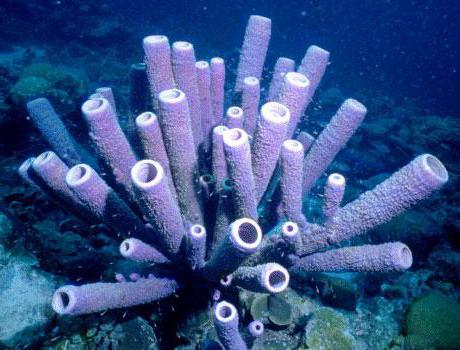
The size of the sponges varies from 1 cm to 2 m, and the color from dull brown to bright purple. The body shape also varies. Sponges can look like a plate, a ball, a fan or a vase.

Nutrition
According to their feeding method, representatives of the Sponge type are heterotrophic filter feeders. Water continuously moves through their body cavity. Thanks to the activity of flagellar cells, it enters the pores of the body layers, enters the atrial cavity and exits through the orifice.
In this case, protozoa, bacteria, phytoplankton and the remains of dead organisms are captured by amoebocytes. This happens by phagocytosis - intracellular digestion. Unprocessed food debris again enters the cavity and is thrown out through the mouth.
There are also predators among sponges. They lack a water-bearing filtration system. Their food is small crustaceans and fish fry that stick to their sticky threads. Then they shorten, pulling towards the body of the predator. The sponge envelops the prey and digests it.

Breathing and elimination
Animals belonging to the Sponge phylum are not found on land. Therefore, they are adapted to absorb oxygen only from water. This happens through diffusion. All cells of the sponge body are capable of absorbing oxygen, as well as removing carbon dioxide.

Asexual reproduction
Despite the primitiveness of their structure, the methods of reproduction of sponges are quite diverse. They can reproduce by budding. In this case, a protrusion appears on the animal’s body, which increases in size over time. When all types of cells have formed on such a bud, it is disconnected from the mother and begins to exist independently.
The next method of sponge reproduction is fragmentation. As a result, the body of the sponge is divided into parts, each of which gives rise to a new organism. This process is also called gemulogenesis. It usually occurs with the onset of unfavorable conditions.
The resulting parts of sponges are called gemmules. Each of them is covered with a protective shell, and inside contains a supply of nutrients. Gemmules are considered the resting stages of sponges. Their ability to survive is simply incredible. They remain viable after exposure to low temperatures down to -100 degrees and prolonged dehydration.
Sexual reproduction
The reproductive process is carried out by specialized cells. In this case, the sperm leaves the mouth of one sponge and flows with a stream of water to the other. There, amebocytes deliver it to the egg.
According to the type of development, sponges are distinguished between ovoparous and viviparous. In the former, the division of the fertilized egg and the formation of the larva occurs outside the mother's body. Such organisms are always dioecious. Among viviparous representatives, hermaphrodites are often found. In them, the development of the zygote occurs in the atrial cavity.

Ecology
For the spread of animals such as Sponges, the presence of a certain substrate is of great importance. It must be firm because silt can clog in the pores. This leads to mass death of animals.
Characteristics of the Sponge type would be incomplete without mention of symbiosis. In nature, there are known cases of their mutually beneficial cohabitation with other aquatic inhabitants. It could be algae, bacteria or fungi.
With this form of existence, the metabolism of sponges occurs more intensively. For example, when cohabiting with algae, they release several times more oxygen and organic matter. Since adult sponges are inedible, many animals use them to protect themselves from enemies. There are cases when crustaceans settle in them. And crabs prefer to wear sponges on their shells.

Meaning in nature and human life
Sponges are of great importance for cleaning water bodies. By filtering, they not only feed, but also remove contaminants. These animals also play their role in trophic chains. Molluscs and certain types of fish feed on sponge larvae.
For humans, sponges are raw materials for pharmacology. Everyone knows sponge-based ointments for bruises and contusions - badyagi, as well as iodine-containing medications. The meaning of these animals is also connected with their name. They have been used for a really long time to wash the body and various surfaces. And now we call such synthetic products sponges.
So, in the article we looked at representatives of the subkingdom Multicellular - the Sponge type. These are multicellular aquatic animals that lead an attached lifestyle. There are two layers in their body - ecto- and endoderm. Each of them is formed by specialized cells. Sponges do not form real tissues.
